Introduction
The IEEE EMBS International Summer School of Neural Engineering (ISSNE) is a platform that has biennially gathered researchers, scientists, and students in the neural engineering field since 2013. The 6th ISSNE, scheduled for July 29 to August 3, 2024, will spotlight the theme of "AI-Empowered Brain Imaging & Decoding." This year's program is tailored to delve into the overlap of artificial intelligence and neuroimaging & neural coding, underscoring the transformative influence of AI within this area. ISSNE will host a series of keynote and seminar talks from distinguished scientists, offering invaluable insights into cutting-edge developments and trends in AI-based brain imaging and decoding. Students will also benefit from a bootcamp training specifically designed for AI-based discovery, providing them with tangible skills and knowledge. Plus, the 2024 ISSNE will facilitate student poster and slide presentations. This will allow participants to exhibit their innovative research findings and ideas, with the potential to win awards for presentations. We invite you to join us at the 2024 ISSNE and immerse yourself in the thrilling exploration of AI-based brain imaging and decoding research.
IEEE EMBS国际神经工程暑期学校(ISSNE)自2013年以来,每两年一届,汇聚神经工程领域的研究人员、科学家和学生。第六届ISSNE定于2024年7月29日至8月3日举行,将聚焦“AI赋能的大脑成像与解码”这一主题。今年的项目旨在深入探讨人工智能与神经成像和神经编码的交叉领域,强调AI在该领域的变革性影响。ISSNE将举办一系列由杰出科学家主讲的主题演讲和专题报告,提供基于AI的大脑成像和解码领域的前沿发展和趋势的宝贵见解。学生还将受益于专为AI基础探索设计的训练营培训,获得实践技能和知识。此外,2024年ISSNE将举办学生海报和口头报告展示,让参与者展示他们的创新研究成果和想法,并有机会获得演讲奖。我们诚挚地邀请您参加2024年ISSNE,遨游在基于AI的大脑成像和解码研究中。
Program Outline
|
Day 0: Registration and ICE Breaking |
||||
|
Day 1 (07/30) |
Day 2 (07/31) |
Day 3 (08/01) |
Day 4 (08/02) |
Day 5 (08/03) |
|
Opening 30 min Keynote 1 Anqi Qiu Keynote 2 Bo Hong |
Keynote 3 Ching-Po Lin Invited Session Talk 1,2 Ching-Po Lin Xia Wu |
Keynote 4 Parashkev Nachev Invited Session Talk 3,4 Ze Wang Yao Li |
Keynote 5 Zhi-Pei Liang Invited Session Talk 5,6 Yao Li Xiaoyong Zhang |
Keynote 6 Dan Wu Invited Session Talk 7,8 Dan Wu Yu Sun |
|
BootCamp Training 1 Anqi Qiu |
Campus Tour (Xiaoli Guo) |
BootCamp Training 2 (Shanbao Tong) |
Industrial Tour (Yue Guan) |
Student Presentations Closure |

Affiliation: School of Biomedical Engineering, Tsinghua University, China
Dr. Bo Hong received his Ph.D. degree of Biomedical Engineering from Tsinghua University in 2001. From 2004 to 2005, he was a visiting scientist in the Department of Biomedical Engineering and the Center for Neural Engineering at Johns Hopkins University, USA. He is now full professor with School of Biomedical Engineering, Tsinghua University, and an investigator of McGovern Institute for Brain Research at Tsinghua. His main research interests are brain computer interface and language network in human brain. His team designed and developed minimally invasive BCI – NEO system and conducted the first-in-human clinical trial successfully in 2023. He has co-authored more than 80 papers on Nature Neuroscience, PNAS, Nature Communications, NeuroImage, Journal of Neuroscience etc. He has served as the Associate Editor of IEEE Transactions on Biomedical Engineering and IEEE Transactions on Neural Systems and Rehabilitation Engineering.
Title: Minimally Invasive Brain Computer Interface: From Bench to Bed (微创脑机接口:从实验室到临床)
Abstract: Intracranial BCI can assist severely disabled persons in assistive communication and active rehabilitation. Nevertheless, sustainable BCI implants require minimal invasiveness. We developed an intracranial BCI for spelling, utilizing only three electrodes over the middle temporal visual area (MT), to pick up focal visual motion responses. The best recording electrodes were decided by preoperative fMRI imaging. The BCI spelling system reached an equivalent information transfer rate of about 20bits/min per electrode. validated a new approach of balancing the intracranial BCI speed and invasiveness.
Taking this minimally invasive strategy, we developed a miniaturized epidual BCI implant with only 8 electrodes (NEO system). The 25mm-diameter device can be fitted in the skull, with no battery included. Power was supplied remotely through inductive antenna, and the epidual ECoG were transmitted wirelessly to the receiver attached outside the scalp. The NEO system was tested on white pigs, showing its capacity of stable long-term recording of epidual ECoG, while keeping cortical cells intact. With ensured minimal invasiveness and long-term safety, the first-in-human trial of clinical implantation was made on October 24th, 2023, in Beijing Xuanwu Hospital, and the second on December 19 th, 2023 in Tiantan Hospital respectively. The first tetraplegia patient with spinal cord injury regained the ability of grasping objects, and even drinking water with help of the BCI. The second BCI-implant patient can control a computer cursor to play games and maneuver a wheelchair just by motor imagery. Both patients have been using the NEO BCI at their home for over six months
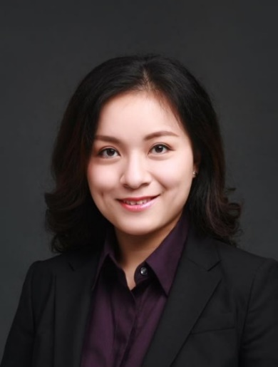
Affiliation: School of Biomedical Engineering, Shanghai Jiao Tong University, China
Yao Li received her bachelor degree from Shanghai Jiao Tong University and her Ph.D. degree from State University of New York at Stony Brook. She is currently the Professor at the School of Biomedical Engineering and serves as the associate dean of National Engineering Research Center of Advanced Magnetic Resonance Technology at Shanghai Jiao Tong University. Dr. Li’s research is in the general area of magnetic resonance spectroscopic imaging, multi-molecular brain mapping, and multi-modal brain imaging for brain disorders. Her work has been recognized by a number of awards, including the W. S. Moore Award (ISMRM, 2023), Eastern Scholar Award (2023), IEEE-EMBC Best Paper Award (2021), Guangci Scholar Award (2021), Summa Cum Laude Award (ISMRM, 2019-2024), Magna Cum Laude Award (ISMRM, 2019-2023). She has authored over 100 papers in journals such as Science, Brain, and Magnetic Resonance in Medicine. Dr. Li currently serves as an Associate Editor for BMC Neuroscience journal. She is a Senior Member of IEEE and a Lifetime Member of the World Chinese Biomedical Engineering Society (WACBE). She was nominated as a member of the Administrative Committee of IEEE EMBS in 2020.
Title: Intelligent MR Image Segmentation of the Brain (智能磁共振脑影像分割)
Abstract: Accurate segmentation of brain tissues or brain lesions is essential for the treatment planning of brain disorders. Learning the high-dimensional spatial-intensity distributions of brain images poses a significant challenge for both classical model-based and deep learning-based methods. In this talk, I will introduce a new framework for learning the spatial-intensity distributions of normal and lesion brain tissues by synergistically integrates brain tissue spatial priors in the form of a subspace with deep generative model. The subspace-based generative model effectively captures both global and local spatial-intensity distributions that are position-dependent. This proposed framework can be adaptively applied to brain tissue and lesion segmentation, achieving significantly improved performance in both accuracy and robustness compared with state-of-the-art methods.
Title: Towards Unraveling the Structural, Functional & Molecular Fingerprints of Brain Function and Disorders (脑功能和脑疾病结构、功能及分子指纹探究)
Abstract: Brain mapping is one of the most exciting frontiers of contemporary science. It provides this generation of scientists the opportunity to make major advances on a historic question: how the brain works and what goes wrong when it is injured or diseased. Magnetic resonance (MR)-based neuroimaging techniques have been widely used for brain research because of their noninvasiveness and multimodal capabilities. Exploiting the rich information contents of MR signals, we can obtain structural, functional and metabolic information of the brain. MR spectroscopic imaging (MRSI) is a beautiful integration of MR spectroscopy and MR imaging, which can provide metabolic status of tissues in vivo without exogenous molecular labels. But its clinical applications have been limited due to long scan time and poor spatial resolution. In this talk, I will introduce a novel capability for rapid high-resolution metabolic imaging using a recently developed subspace-based imaging technique called SPICE (Spectroscopic Imaging by Exploiting Spatiospectral Correlation). With approximately 10 minutes of scanning time, we can acquire metabolic maps of the entire brain at a nominal spatial resolution of 2.0 x 3.0 x 3.0 mm³. We have successfully conducted SPICE scans on patients with stroke, Alzheimer's disease (AD), multiple sclerosis (MS), and brain tumors. This lecture will briefly cover the clinical studies and current findings in multi-molecular imaging of the brain using SPICE, which have provided significant insights into the pathophysiology of brain disorders in clinical settings. By leveraging the unique strengths of multimodal and multi-molecular brain MR imaging, we can explore the interlinked relationships among different pathological biomarkers and improve the diagnostic and prognostic performance for complex brain disorders.

Affiliation: Institute of Neuroscience, Yang Ming Chiao Tung University, Taiwan; Department of Education and Research, Taipei City Hospital, Taiwan
Prof. Ching-Po Lin serves as the Head of the MRI Core at National Yang Ming Chiao Tung University, overseeing facilities that include a 3T Human MRI (Prisma, Siemens) and a 7T Preclinical PET/MRI (Biospec, Bruker). He also serves as the Director of the Education and Research Department at Taipei City Hospital and holds the position of Adjunct Investigator at the Institute of Biomedical Engineering and Nanomedicine at the National Health Research Institutes in Taiwan. His research primarily focuses on developing innovative methodologies for imaging human brain connectomics. A pivotal aspect of his work is ensuring the accuracy of neural tracts mapping, a critical component for advancing precision medicine and deepening our understanding of brain functions and disorders. His efforts are dedicated to developing, validating, optimizing, and applying these methodologies to explore the brain networks that underpin cognitive performance and their alterations due to disconnection syndromes, such as aging, dementia, depression, and schizophrenia. By measuring changes in connectivity, he has identified early markers of aging and dementia, establishing links between brain networks and behavioral as well as clinical indices. More recently, he has committed to integrating multimodal MRI techniques to enhance preoperative planning for brain surgeries, improve intraoperative navigation, and refine postoperative evaluations in patients with epilepsy and brain tumors. To date, he has secured 12 invention patents and authored 282 SCI papers, amassing over 17,800 citations with an H-index of 59.
Keynote:
Title: Mapping the connectomics of human brain disorders (人类大脑疾病的连接组学图谱绘制)
Abstract: The human brain comprises a complex neural network, known as the human connectome, with a broad range of regional microscale cellular morphologies and macroscale global properties, forming an efficient system for processing and integrating multimodal information. Plenty of evidence has shown that mapping the connections may be beneficial to understand normal variability as well as mental disorders. It may also be crucial for pursuing a new generation algorithm of artificial intelligence and thus has the so-called 21st-century brain.
Due to the limited available techniques, neuroscientists exhibited little interest in the connections for centuries till modern non-invasive neuroimaging techniques were invented in the 1990s. The structural connections, as well as related neural activities, thus attracted people’s attention and thus opened the prolog of the human brain connectome. Opportunely, Prof. Lin caught up with the tide for his Ph.D. thesis and founded the Brain Connectivity Lab, dedicated to driving modern neuroimaging methods for brain connectomics studies, after moving to Yang Ming University in 2004.
By studying the direction-dependent water molecular diffusivity and blood oxygen level coherence, brain structural and functional connectivity could be mapped through modern MRI techniques, thus opening a window to explore functional anatomy based on its structural substrates. Meanwhile, the complexity of higher brain systems emerged as another challenge to explain the rich functionalities that arise from a relatively fixed structure. To facilitate human brain study, we are dedicated to driving technical developments and have striven to optimize these methods under human brain study regulations for decades. Accordingly, reliable and efficient neuroimaging protocols were established to promise further brain connectomics studies, including clarifying human brain functions and assisting clinical services, including neurosurgical plans, neurodegenerative prediction, and biosignatures for psychiatric disorders. Hereby, this lecture will discuss modern imaging technologies for human connectome mapping and the recent progress in related works.
Session talk:
Title: Leveraging MRI and AI to Forecast Brain Aging (利用MRI和人工智能预测脑龄)
Abstract: Brain aging is a multifaceted process shaped by an array of genetic, environmental, and lifestyle elements. Traditional methods for estimating brain age have relied on subjective evaluation, often lacking in precision. Currently, through the application of MRI technology, researchers can acquire comprehensive structural and functional insights into the complex operations of the human brain. In conjunction with advanced AI algorithms, such as machine learning and deep learning, and extensive MRI datasets, researchers have crafted predictive models that yield precise brain age estimates.
In this presentation, we will delve into the technical intricacies of harnessing MRI and AI for brain age prediction, capitalizing on the remarkable progress achieved over the past decade. We will scrutinize various MRI techniques and analytical methods employed to pinpoint critical biomarkers of brain aging. Additionally, we will explore the ramifications of brain age prediction in both clinical and research environments.
Accurate brain age estimation has substantial potential for gauging cognitive health, identifying age-related disorders, and assessing the efficacy of therapeutic interventions. It paves the way for tailored healthcare strategies, early identification of neurological disorders, enhanced diagnostic precision, and the encouragement of healthy aging processes in the brain. This burgeoning field also offers a hopeful avenue for unraveling the complexities of brain aging, fostering innovative approaches to personalized care, and proactive management of brain health.

Affiliation: ECE Department of Electrical and Computer Engineering, University of Illinois at Urbana-Champaign, United States
Zhi-Pei Liang received his Ph.D. degree from Case Western Reserve University in 1989. He subsequently joined the University of Illinois at Urbana-Champaign (UIUC) first as a postdoctoral fellow and then as a faculty member. Dr. Liang is currently the Franklin W. Woeltge Professor of Electrical and Computer Engineering; he also chairs the Computational Imaging Group in the Beckman Institute. Dr. Liang’s research is in the general area of magnetic resonance imaging, signal processing and machine learning. His work has been recognized by a number of awards, including the Sylvia Sorkin Greenfield Award (Medical Physics, 1990), the Whitaker Biomedical Engineering Research Award (1991), the NSF CAREER Award (1995), the University Scholar Award (UIUC, 2001), the Isidor I. Rabi Award (ISMRM, 2009), the Otto Schmitt Award (IFMBE, 2012), IEEE-EMBC Best Paper Awards (2010, 2011, 2021), IEEE-ISBI Best Paper Awards (2010, 2015), the Technical Achievement Award (IEEE-EMBS, 2014), and the Gold Medal (ISMRM, 2022). Dr. Liang was selected as the Paul C. Lauterbur Lecturer for the 2016 ISMRM meeting and as the Savio L. Woo Distinguished Lecturer for the 2017 WACBE World Congress on Bioengineering. He is a Fellow of IEEE, ISMRM and AIMBE. He was elected to the International Academy of Medical and Biological Engineering in 2012 and to the US National Academy of Inventors in 2021. Dr. Liang served as President of the IEEE-EMBS 2011-2012 and received its Distinguished Service Award in 2015. He has been serving as Chair-elect of the International Academy of Medical and Biological Engineering since 2022.
Title:SPICEx: Towards an Omni Imaging Technology for Brain Mapping (SPICEx:全能脑成像技术)
Abstract:
Scientific research requires powerful tools for elucidating the secrets of nature. Today, understanding the brain — how it works and what goes wrong when it is injured or diseased, is considered one of the last frontiers in science, and it has posed significant challenges for scientists and engineers to develop effective tools to meet this objective. This talk will discuss a new AI-powered magnetic resonance (MR) spectroscopic imaging technology, known as SPICEx, for ultrafast, high-resolution, labeled-free molecular imaging of the brain. SPICEx uses a machine learning framework to effectively integrate quantum simulation, rapid scanning, sparse sampling, and constrained image processing to provide an unprecedented capability for simultaneous mapping of brain structures, function and metabolism using tissue intrinsic MR spectroscopic signals from multiple molecules. In this talk, I’ll will give an overview of SPICEx, show some “SPICY” experimental results, and discuss future opportunities of AI-powered brain mapping.

Affiliation: Institute of Neurology, University College London, United Kingdom
Parashkev is a neurologist and neuroscientist. He has been an early advocate for the use of complex modelling in understanding brain and behaviour, demonstrating the necessity for a high-dimensional approach to the domain in advance of its recent technological realisation. His High-Dimensional Neurology Group at the UCL Queen Square Institute of Neurology is focused on the development of a comprehensive computational framework for drawing predictive, inferential, and prescriptive intelligence from large-scale, real-world, multimodal clinical data, innovating across the methodological domains of brain registration, featurization, representation, and topological and causal inference, and the modelling of cognition and behaviour with disruptive data.
Title:Foundation modelling of the human brain (人类大脑基础建模)
Abstract: Biological systems, the human brain pre-eminently amongst them, must be capable of adapting to highly complex environments. Since any good controller can be no simpler than its target, this sets a high bound on their minimal organisational complexity. Understanding the physiology of the brain--and prescribing its manipulation in disease--will therefore require far more flexible models than neuroscience currently employs. The critical question is how to approach the task given real-world constraints on available data, compute, and algorithmic ingenuity. Here I argue that multi-modal deep generative modelling offers the only plausible solution, and examine the challenges and potential rewards of applying this emerging technology to the imaged brain, drawing on analyses involving >10^6 individual brain scans across >10^5 distinct patients powered by >5 petaFLOPS of compute, in the context of an array of representational, predictive, and prescriptive tasks.
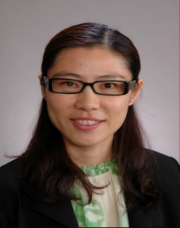
Affiliation: Department of Health Technology and Informatics, the Hong Kong Polytechnic University, Hong Kong
Dr. Qiu is a global STEM scholar and Professor at the Department of Health Technology and Informatics at the Hong Kong Polytechnic University. She is also an Adjunct Professor at the Department of Biomedical Engineering at the Johns Hopkins University. Her past roles include Deputy Head for Research & Enterprises at the Department of Biomedical Engineering and Director for the BME Innovation Center at the NUS Suzhou Research Institute, a part of the National University of Singapore. Dr. Qiu commenced her academic journey with a BS in Biomedical Engineering from Tsinghua University. She then earned two MS degrees - one in Biomedical Engineering from the University of Connecticut, and another in Applied Mathematics and Statistics from Johns Hopkins University. Her continued dedication to academia led her to earn a PhD from Johns Hopkins. Specializing in computational analyses, Dr. Qiu is deeply committed to understanding the origin of individual health differences throughout a lifespan. She leverages complex and informative datasets that include disease phenotypes, neuroimaging, and genetics to further her research. Her team has high-impact publications in Nature, Nature Neuroscience, Nature Mental Health, American Journal of Psychiatry, Biological Psychiatry, IEEE Transactions in Medical Imaging, Medical Image Analysis.
Title: Spectral Laplace-Beltrami Wavelets and Geometric Convolutional Neural Network for Signal Processing and Classification (谱拉普拉斯-贝尔特拉米小波与几何卷积神经网络在信号处理与分类中的应用)
Abstract:
The Laplace-Beltrami operator is a generalization of the Euclidean representation of the Laplace operator to an arbitrary Riemannian manifold. It is a self-adjoint operator and its eigenfunctions form a complete set of real-valued orthonormal basis functions. In this talk, I will introduce spectral Laplace-Beltrami wavelets and its computational algorithm. I will then demonstrate its use for smoothing and classification of the data defined on smooth surfaces embedded in the 3-D Euclidean space. Furthermore, I will discuss that the spectral Laplace-Beltrami Wavelets and Hodge-Laplacian to incorporate graph network topology can be used for the construction of geometric convolutional neural network (CNN). I will show the use of this method on brain morphology and functional networks for the prediction of Alzheimer’s Disease and Cognition in adolescents.
Bootcamp Title: Practice on Graph Neural Network for Brain Data (图神经网络处理大脑数据实战)
Bio for Teaching assistant for the Bootcamp:
Jinghan Huang received his BS degree from the School of Physics and Astronomy at Shanghai Jiao Tong University, China, and his MEng degree in Biomedical Engineering from the National University of Singapore. He is working towards a PhD in Computer Science at the University of Manchester. His current research interests include graph deep learning and its applications to medical image analysis.
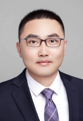
Affiliation: College of Biomedical Engineering and Instrument Science, Zhejiang University, China
Prof. Yu SUN is a tenured associate professor at Zhejiang University with expertise in neural engineering and brain-computer interface. He has published more than 100 papers (including 10 papers with EMBS Best paper award, ESI highly-cited, cover and featured articles). Prof. SUN was recognized as the World Top 2% Scientists (BME) by Stanford University, Hanxiang Young Scientist by the EEG-related Technology Committee, Chinese Psychological Society, Outstanding Young Scholar on the Frontier of Chinese Engineering by the Chinese Academy of Engineering, Distinguished Expert of Zhejiang Province. Currently, he serves as the editor board of IEEE JBHI, TNSRE, MBEC.
Title: Passive Brain-computer Interface: New frontier of neural engineering(被动式脑机接口:神经工程前沿)
Abstract: In recent years, unlike traditional active brain-computer interface (BCI) technologies that primarily focus on the recognition of subjective intentions and interaction with internal and external environments, passive BCI technologies aimed at brain state detection have gradually emerged as a new research hotspot in the field of brain-computer interfaces. Passive BCI technologies open a window to objectively, in real-time, and continuously monitor cognitive and emotional states of the brain, enabling researchers to more intuitively evaluate the complex psychological states of subjects. This has significant implications for professionals working in highly specialized environments

Affiliation: School of Biomedical Engineering, Shanghai Jiao Tong University, China
Dr. Shanbao Tong received a B.S. in radio technology from Xi'an Jiao Tong University (Xi'an, China) in 1995, an M.S. degree in turbine machine engineering, and a Ph.D. in biomedical engineering from Shanghai Jiao Tong University in 1998 and 2002, respectively. From 2000-2001, he was a research trainee at the Johns Hopkins School of Medicine. From 2002-2005, he had postdoctoral training at the Johns Hopkins School of Medicine. Dr. Tong is the founding chair of the IEEE EMBS Shanghai chapter. He is also the founding chair of the IEEE EMBS International Summer School of Neural Engineering (ISSNE). He was the associate editor of IEEE Transactions on Biomedical Engineering and IEEE Transactions on Neural Systems and Rehabilitation Engineering and the conference chair of the 2017 IEEE EMBS International Conference of Neural Engineering (NER'17). He is currently the editor-in-chief of Medical & Biological Engineering & Computation.
Bootcamp Title: Statistic fallacies and data leakages in biomedical data analysis (生物医学数据分析中的统计谬误和数据泄漏问题)
Abstract: Biomedical signal and image analysis are crucial for biomedical research and clinical studies. However, there is a reproducibility crisis in biomedical research due to a lack of rigorous data analysis. As part of this year's IEEE EMBS International Summer School of Neural Engineering, we will be hosting a half-day, three-hour bootcamp titled "Statistic Fallacies and Data Leakages in Biomedical Data Analysis." This bootcamp will cover common statistical fallacies and data leakage problems in biomedical data analysis, including
· data leakage issues in machine learning,
· misinterpreting confidence intervals and null hypothesis significance testing,
· p-hacking,
· HARKing (Hypothesizing After the Results are Known), and
· misuses and misinterpretations in correlation analysis
Through case studies and hands-on activities, this bootcamp aims to help students understand common statistical fallacies and data leakage issues, enabling them to conduct more replicable research.
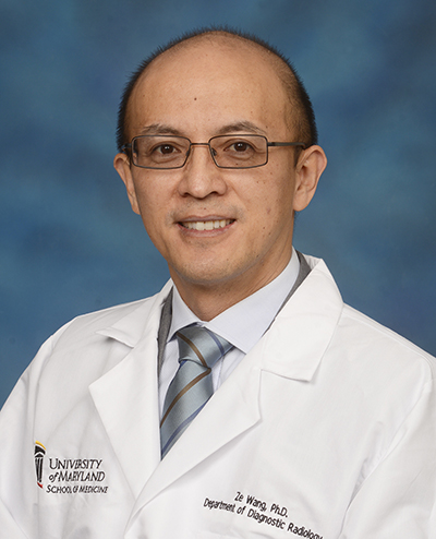
Affiliation: School of Medicine, University of Maryland, United States
Ze Wang, PhD, Tenured Full Professor of Radiology, University of Maryland School of Medicine. Dr. Wang got his PhD from Shanghai Jiao Tong University in 2003. Dr. Wang was a Research Assistant Professor at University of Pennsylvania from 2009-2014, and a Professor in Hangzhou Normal University from 2014-2016. He joined Temple University as an Associate Professor and the Director of MRI Center in 2016. In 2019, he moved to University of Maryland School of Medicine as an Associate Professor. He has published 128 peer-reviewed papers mostly as the leading author and or senior author. His H-index is 48. Dr. Wang’s research focuses on MR technique development, image processing, biosignal processing, machine learning, neuroimaging in Alzheimer’s Disease and drug addiction. He has developed 4 widely used software packages: 3D FSE spiral readout ASL perfusion MRI sequence and online reconstruction program, ASL signal processing toolbox (ASLtbx), Brain entropy mapping toolbox (BENtbx), multivariate-lesion symptom mapping toolbox (SVR-LSM). Dr. Wang received the Shanghai Excellent Thesis Award, and the China National Top 100 PhD Theses Nomination Award, both in 2005. He is an editorial board member or associate editors for several international journals and an ad hoc reviewer for NIH and other international foundations. He currently leads three NIH R01 grants, a R21 project, a R21/R33 project as the PI. He is also supported by a P41 sub-contract and an internal seed fund. More information can be found in https://www.medschool.umaryland.edu/pi/Ze-Wang-PhD/
Title: Machine Learning in Medical Imaging (机器学习在医学影像中的应用研究)
Abstract:
In this lecture, I will provide a brief overview of machine learning in medical imaging including data acquisition, reconstruction, and processing. I will mainly focus on MRI. To be self-contained, brief introduction about imaging will be provided as well. For machine learning, a major focus will be supervised machine learning. Learning points in my talk will include: the rationale for why deep learning can solve highly complex technique problems; capability of deep learning for denoising and data acquisition acceleration.
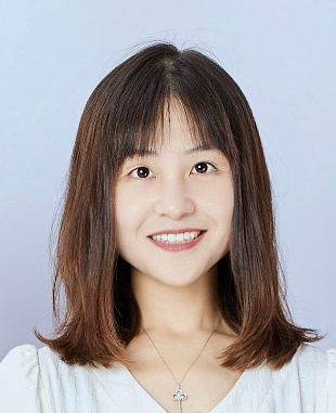
Affiliation: College of Biomedical Engineering and Instrument Science, Zhejiang University, China
Dan Wu is currently a Professor at Zhejiang University. Dr. Wu obtained her Bachelor’s degree from Zhejiang University and master and PhD degrees from the Department of Biomedical Engineering at Johns Hopkins University. She served as an Assistant Professor at the Department of Radiology, Johns Hopkins University School of Medicine.
Dr. Wu’s research focuses on the development of MRI pulse sequence and neuroimage analysis techniques, with a focus on diffusion MRI and developmental neuroscience. Dr. Wu has published over 100 peer-reviewed journal articles on PNAS, Science Advances, Radiology, etc. She is the PI of over 10 national and provincial projects. She was awarded Innovator under 35 China by MIT Tech Review, Young Scientist of World Economic Forum, Excellent Youth Project of NSFC, etc. Dr. Wu served the Associate Editor of Human Brain Mapping, and the Chair of Pediatric MR Study Group and the Secretary of the Diffusion MR Study Group of the International Society of Magnetic Resonance in Medicine (ISMRM).
Keynote talk
Title: Mapping the brain connectome from macro-, meso-, and microscopic MRI (基于宏观、介观和微观MRI绘制大脑连接组图谱)
Abstract: Recent development of diffusion MRI has enabled non-invasive mapping of tissue microstructures, and has been widely used in clinical diagnosis and neuroscience. This talk will first introduce the basics of diffusion MRI, and then concept and methodology of diffusion MRI-based microstructural imaging. Particularly, we introduce our recent effort in developing of time-dependent diffusion MRI, and its applications in tumors and brain disorders, as well as our recent effort on developing an ultra-high-gradient system to maximize the potential of microstructural imaging.
Session talk
Title: Spatiotemporal atlas of the developing brain (发育中大脑的时空图谱)
Abstract: Neuroimaging of the developing, despite its technical challenge, has been shown to be important to tool to visualize the brain development and to assistant diagnosis of developmental diseases. This talk will cover our recent endeavors to develop advanced imaging techniques for fetal and infant brain MRI, and the generation of spatiotemporal atlas of early brain development, as well as the large data-based neuroimaging analysis of the child and adolescent brains.

Affiliation: School of Computer Science, Beijing Institute of Technology, China
Xia Wu is the professor of the School of Computer Science & Technology, Beijing Institute of Technology, mainly studying the intersection of AI and brain science. She has published more than 100 high-level articles as the first/corresponding author in journals and top international conferences. Prof. Wu has received a grant from the National Science Fund for Distinguished Young Scholars, the First Prize in Wu Wenjun AI Science and Technology Award for Natural Science, and the Science Research Famous Achievement Award in Higher Institution from the Ministry of Education, the Mao Yi-sheng Science and Technology Award - Beijing Youth Science and Technology Award.
Title: When the Brain Encounters AI: The Development from Neural Decoding to General AI
(当大脑遇上AI:从神经解码到通用AI的发展)
Abstract:
The human brain is the core of human intelligence, and the study of the brain, especially the mechanisms of higher cognitive functions, has become an important research topic. With the development of computer technology, neuroscientists have used machine learning and artificial intelligence algorithms to analyze brain activity, providing the possibility to reveal the mysteries of internal brain activity. This report starts from the development, application, methodology, challenges, and future directions of brain decoding technology, introducing the application of advanced intelligent analysis algorithms in brain decoding, with the aim of helping to further clarify the brain's neural mechanisms and promote the development of brain science. At the same time, the clarification of brain mechanisms will also provide theoretical support and inspiration for the development of general artificial intelligence.

Affiliation: School of Medicine,Shanghai Jiao Tong University
Xiao-Yong Zhang is a principal investigator at Shanghai Jiao Tong University School of Medicine. He is dedicated to developing magnetic resonance imaging (MRI) technology and artificial intelligence algorithms for healthcare. Specifically, he focuses on: 1) developing MRI techniques to visualize the microenvironment of brain diseases, understand their heterogeneity, and advance disease diagnosis; 2) developing deep learning algorithms to integrate multi-scale medical data for disease prevention, diagnosis, prognosis, and treatment design. To date, he has published more than 60 journal papers, with representative works appearing in journals such as "Medical Image Analysis," "IEEE Transactions on Medical Imaging," and "Advanced Science." He holds positions as a standing committee member or member of several national-level academic societies and serves as an editorial board member for journals.
Title: Developing intelligent diagnosis and image generation models for brain diseases based on a single MRI modality (基于单一MRI模态的智能诊断和图像生成模型在大脑疾病中的应用)
Abstract:
Magnetic Resonance Imaging (MRI) is a powerful non-invasive tool used in the diagnosis of brain diseases. While multimodal MRI provides comprehensive insights, single MRI modality remains highly relevant due to its accessibility and reduced complexity. Our work focuses on developing intelligent models for the diagnosis and image generation of brain diseases using data derived from a single MRI modality. Leveraging advanced machine learning and deep learning algorithms, we aim to enhance the accuracy of diagnosis and the quality of image synthesis. Our proposed artificial intelligence (AI) models integrate state-of-the-art methodologies to identify and segment pathological features in MRI scans, facilitating precise disease characterization. Additionally, we explore image generation techniques that simulate high-quality brain images, potentially aiding in the augmentation of limited datasets and providing valuable synthetic data for training and validation purposes. The results of our work demonstrate the potential of AI-driven approaches in improving diagnostic accuracy and generating realistic MRI images, offering significant benefits for clinical applications and research in neuroscience.
Application & Registration
1. Please submit your application through the link , or scan the QR code:

2. For any inquiries, please contact Ms. Hillary Shang at: issne2024@sjtu.edu.cn
3. Registration will be starting from June 1st, 2024, and the summer school will accommodate a maximum of 100 students.
Preliminary Program
(may subject to minor change, updated on June 11)
Day 1 (July 30)
Keynote 1:
Anqiu Qiu, Spectral Laplace-Beltrami Wavelets and Geometric Convolutional Neural Network for Signal Processing and Classification (谱拉普拉斯-贝尔特拉米小波与几何卷积神经网络在信号处理与分类中的应用)
Keynote 2:
Bo Hong, Minimally Invasive Brain Computer Interface: From Bench to Bed (微创脑机接口:从实验室到临床)
BootCamp Training 1:
Anqi Qiu,
Practice on Graph Neural Network for Brain Data (图神经网络处理大脑数据实战)
Day 2 (July 31)
Keynote 3:
Ching-Po Lin, Mapping the connectomics of human brain disorders (人类大脑疾病的连接组学图谱绘制)
Invite Session Talk 1, 2
Ching-Po Lin: TBD
Xia Wu, TBD
Campus Tour
Day 3 (Aug 1)
Keynote 4:
Parashkev Nachev, Foundation modelling of the human brain (人类大脑基础建模)
Invited Session Talk 3,4
Ze Wang, Machine Learning in Medical Imaging (机器学习在医学影像中的应用研究)
Yao Li, Intelligent MR Image Segmentation of the Brain (智能磁共振脑影像分割)
BootCamp Training 2:
Shanbao Tong, Statistic fallacies and data leakages in biomedical data analysis (生物医学数据分析中的统计谬误和数据泄漏问题)
Day 4 (Aug 2)
Keynote 5:
Zhi-Pei Liang, SPICEx: Towards an Omni Imaging Technology for Brain Mapping (SPICEx:全能脑成像技术)
Invited Session Talk 5,6
Yao Li, Towards Unraveling the Structural, Functional & Molecular Fingerprints of Brain Function and Disorders (脑功能和脑疾病结构、功能及分子指纹探究)
Xiaoyong Zhang, TBD
Industrial Tour
Day 5 (Aug 3)
Keynote 6
Dan Wu, Mapping the brain connectome from macro-, meso-, and microscopic MRI (基于宏观、介观和微观MRI绘制大脑连接组图谱)
Invited Session Talk 7,8
Dan Wu, Spatiotemporal atlas of the developing brain (发育中大脑的时空图谱)
Yu Sun, Passive Brain-computer Interface: New frontier of neural engineering(被动式脑机接口:神经工程前沿)
Student Presentations
Closure